The serous membrane is a vital part of your body’s structure, offering both protection and functionality to various organs. This dual-layered membrane secretes serous fluid, facilitating the frictionless movement between tissue surfaces. You can find serous membranes in several key locations within your body, serving as linings for cavities that don’t open directly to the outside.
Examples of serous membranes include the pericardium, which encases your heart; the pleura, surrounding your lungs; and the peritoneum, which encompasses the abdominal cavity. Each of these membranes plays a pivotal role in the smooth operation of your body’s systems, providing lubrication to prevent friction and potential damage during organ movement.
These examples illustrate the serous membrane’s essential contribution to maintaining your physiological integrity.
Basic Structure of Serous Membranes
Serous membranes are vital components in your body that line cavities not open to the outside environment. They possess a simple structure, with two distinct layers that serve both protective and functional roles:
- Parietal Layer: This outer layer lines the internal surface of the body cavity.
- Visceral Layer: In contrast, the inner layer covers the external surface of your organs.
These layers are composed of a single layer of flat, squamous epithelial cells known as mesothelium. Underneath the mesothelium, there is a thin layer of areolar connective tissue.
In between the parietal and visceral layers, you’ll find the serous cavity. This space contains a small amount of lubricating serous fluid, which is secreted by the mesothelium. This fluid allows your organs to glide smoothly against adjacent structures during movements such as breathing or digestion.
Here’s how the layers and features are organized:
| Feature | Description |
|---|---|
| Parietal Layer | Lines the body cavity |
| Visceral Layer | Covers organs |
| Serous Fluid | Fills the space between layers for lubrication |
Some examples of serous membranes include the pleura in the thoracic cavity, the peritoneum in the abdominal cavity, and the pericardium around the heart. Each of these plays a critical role in reducing friction and providing cushioning for your internal organs.
Major Types of Serous Membranes
Serous membranes are pivotal in providing a frictionless environment for vital organs. They line body cavities and organs, ensuring smooth motion and minimizing risk of injury.
Pleura
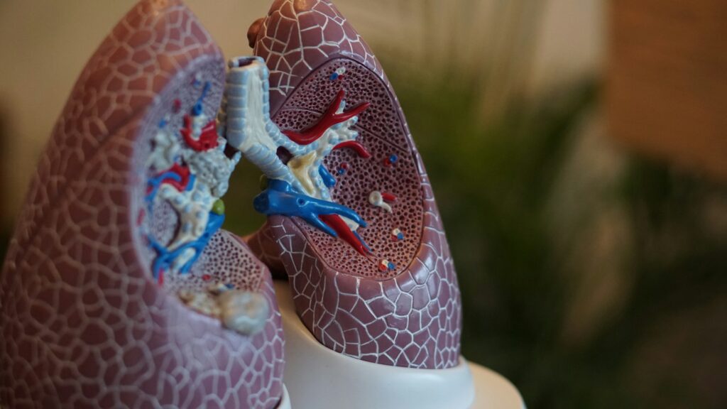
The pleura is the serous membrane enveloping your lungs and lining the thoracic cavity. It consists of two layers: the parietal pleura, lining the thoracic wall, and the visceral pleura, covering the lungs themselves. This membrane produces serous fluid, which aids in effortless lung expansion and contraction during respiration.
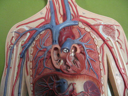
Pericardium
Your heart is encased in the pericardium, another serous membrane, which forms a fluid-filled sac providing a protective cushion. Similar to the pleura, the pericardium features a visceral layer that directly covers the heart and a parietal layer that forms the outer sac, both of which work in tandem to reduce friction as the heart beats.
Peritoneum
The peritoneum is the largest of the serous membranes, lining your abdominal and pelvic cavities. It has two main components: the parietal peritoneum attached to the abdominal wall and the visceral peritoneum wrapping around most of the internal organs.
The peritoneum serves as a lubricant, facilitating the organs’ motions within the abdominal space, which is critical during digestion and other bodily movements.

Pleura Details
The pleura are crucial for your respiratory system, ensuring that your lungs move smoothly within the thoracic cavity during breathing.
Function of Pleura
The primary role of your pleura is to facilitate lung expansion and contraction by providing a lubricated surface. This reduces friction between your lung surface and chest wall as you breathe in and out.
Pleural Layers
There are two main layers of the pleura:
- Visceral pleura: This inner layer directly covers your lungs.
- Parietal pleura: The outer layer that attaches to your chest wall and diaphragm.
These layers create a sealed space known as the pleural cavity, which contains a small amount of fluid enabling the smooth gliding of your lungs during respiration.
Clinical Relevance
The pleura can be affected by various conditions, such as pleuritis or pleural effusion, impacting your lung function. If inflammation or fluid accumulation occurs within your pleural space, it can lead to discomfort or difficulty breathing, necessitating medical intervention. Understanding the anatomy and potential issues with the pleura can be vital for diagnosing and treating thoracic conditions.
Pericardium Characteristics
The pericardium plays a critical role in cardiac function by protecting the heart and maintaining its position within the thorax. Understanding its structure and purpose is key to recognizing its importance in your overall heart health.
Function of Pericardium
The pericardium’s primary function is to encase the heart and provide a smooth, lubricated surface for it to beat without friction. This protective sac ensures the heart moves freely as it pumps blood to your body. Additionally, the pericardium helps to anchor the heart within the chest cavity and prevent its overexpansion.
Pericardial Layers
Your pericardium is composed of two distinct layers: the fibrous pericardium and the serous pericardium. The fibrous layer is a tough, protective shell that safeguards the heart against infections and physical damage.
Within this fibrous component lies the serous pericardium, which includes both a parietal layer lining the fibrous pericardium and a visceral layer directly covering the heart muscle. These serous layers produce a lubricating fluid that reduces friction between the beating heart and surrounding tissues.
- Fibrous pericardium: Dense connective tissue providing durability.
- Serous pericardium: Two-layered structure:
- Parietal layer: Lines the fibrous layer.
- Visceral layer or epicardium: Directly covers the heart.
Cardiac Health Implications
Problems with the pericardium can lead to conditions such as pericarditis (inflammation of the pericardium) or pericardial effusion (excess fluid buildup), which in turn can impact heart function.
Maintaining the integrity and health of the pericardium is essential, as its malfunction can lead to compromised cardiac output and overall cardiovascular health. Regular check-ups and addressing any cardiac symptoms promptly can help safeguard the health of your pericardium.
Peritoneum Aspects
The peritoneum is a pivotal serous membrane in your body, playing essential roles in protecting and structuring the abdominal organs.
Function of Peritoneum
Your peritoneum is not merely a passive structure; it actively facilitates the smooth gliding of abdominal organs against each other. The space it encloses enables the movement and also serves as a conduit for blood vessels, nerves, and lymphatics to pass through.
Peritoneal Layers
There are two main layers of the peritoneum:
- Parietal peritoneum: This layer lines the walls of your abdominal and pelvic cavities.
- Visceral peritoneum: This covers the external surface of most abdominal organs, closely adhering to them.
Between these layers lies the peritoneal cavity, filled with a small amount of fluid that aids in lubrication.
Abdominal Cavity Conditions
Different conditions can affect the peritoneum, such as:
- Peritonitis: Inflammation of the peritoneum, often due to infection or rupture of abdominal organs.
- Ascites: The accumulation of fluid in the peritoneal cavity, which may be caused by various diseases, including liver cirrhosis or heart failure.
Frequently Asked Questions
Serous membranes play a critical role in your body by lining and protecting internal cavities and organs. This section addresses common inquiries regarding their function, structure, and types.
What is the role of serous membranes in the body?
Serous membranes serve to reduce friction, enabling organs to glide smoothly against one another or the walls of cavities during your normal movements and bodily functions. They achieve this by secreting a lubricating serous fluid.
What types of cells are found in serous membrane tissue?
Serous membrane tissue consists of a layer of simple squamous epithelial cells known as mesothelium. These cells are supported by a thin layer of connective tissue and are specialized for fluid secretion.
How does serous fluid facilitate organ function?
Serous fluid acts as a lubricant, which prevents friction between organs as you move. This allows organs, like your heart and lungs, to operate without causing damage to surrounding tissues.
What are the three primary serous membranes within the human body?
The three main serous membranes are the pleura (surrounding the lungs), peritoneum (the largest serous membrane, lining the abdominal cavity), and pericardium (encasing the heart).
Can you explain the difference between parietal and visceral serous membrane layers?
The parietal layer of a serous membrane lines the internal surface of the body cavity, whereas the visceral layer covers the external surface of the organs within that cavity. The space between these layers is where serous fluid is found.
In what ways do serous membranes differ from mucous membranes?
Unlike serous membranes, mucous membranes are associated with body passages and cavities that open to the external environment. They produce mucus, which is thicker than serous fluid and serves to protect and moisten the lining of various organs.

What Visiters Had To Say