Creating a model of an animal cell can be a rewarding project that offers a hands-on way to understand the intricate workings of cells, the building blocks of life. By constructing a three-dimensional representation, you become intimately familiar with the various components that make up an animal cell, from the protective cell membrane to the powerhouse known as the mitochondrion. Such projects require not just creativity but also a commitment to represent the complexity of cells in an accessible and accurate manner.
When embarking on animal cell science projects, you will need to carefully select materials that best illustrate the structure and function of each organelle.
For example, you might use clay or playdough to distinguish between the cell’s components based on color and shape, effectively creating a visual map of an animal cell’s anatomy.
This approach not only solidifies your understanding of each part’s role within the cell but also helps in memorizing their relationships and functions.
Understanding these cellular components is crucial, as they perform the vital processes that enable animals, including humans, to survive and thrive.
Whether you’re constructing an edible model using gelatin and candies or assembling a clay diagram, the educational benefits of this type of project extend beyond the classroom, helping you to grasp fundamental biological concepts that underpin much of the life sciences.
Fundamentals of Animal Cell Biology
In exploring the fundamentals of animal cell biology, you’ll understand the primary components that constitute an animal cell: the protective membrane, the complex cytoplasm with its organelles, and the nucleus housing the genetic material.
Cell Membrane Structure and Function
The cell membrane plays a crucial role as the gatekeeper of the cell. It’s composed of a bilayer of phospholipids, with embedded proteins, allowing it to control the movement of substances in and out of the cell. Its semi-permeable nature ensures that essential nutrients enter while waste products are expelled, maintaining the integrity of the cell.
Cytoplasm and Organelles
Floating within the cytoplasm, organelle structures perform vital functions that keep the cell alive. Mitochondria, often referred to as the powerhouse, generate ATP through respiration. The endoplasmic reticulum and Golgi apparatus work in concert to produce and transport proteins. Lysosomes and peroxisomes help in breaking down waste, defending against pathogens, and more.
Nucleus and Genetic Material
At the core of cell functionality is the nucleus, which houses DNA strands wrapped into chromosomes. It controls all activities by dictating protein synthesis, which in turn regulates cell growth, division, and differentiation through gene expression. The nucleolus within the nucleus is particularly important for the assembly of ribosomes.
Microscopy Techniques
Microscopy is a fundamental tool in animal cell science projects, enabling you to examine cellular structures in exquisite detail. These techniques allow you to prepare, stain, and visualize samples for in-depth analysis.
Preparing Cell Slides
Before you can delve into the microscopic world of cells, you need to properly prepare slides. This involves carefully placing a thin slice of your sample onto a glass slide. It’s vital to ensure the tissue is thin enough to allow light to pass through and that it’s free from air bubbles which can obstruct the view.
- Clean your slide and coverslip thoroughly to avoid contaminants.
- Slice your specimen thinly using a sharp blade or microtome.
- Place the sample on the slide and add a drop of water or mounting solution.
Staining for Cell Observation
Staining is crucial to differentiate and highlight various cellular components under the microscope. Specific stains bind to certain cell parts, such as the nucleus or cell membrane, imparting distinct colors.
- Hematoxylin and eosin (H&E) are common stains that respectively highlight the nucleus and cytoplasmic components.
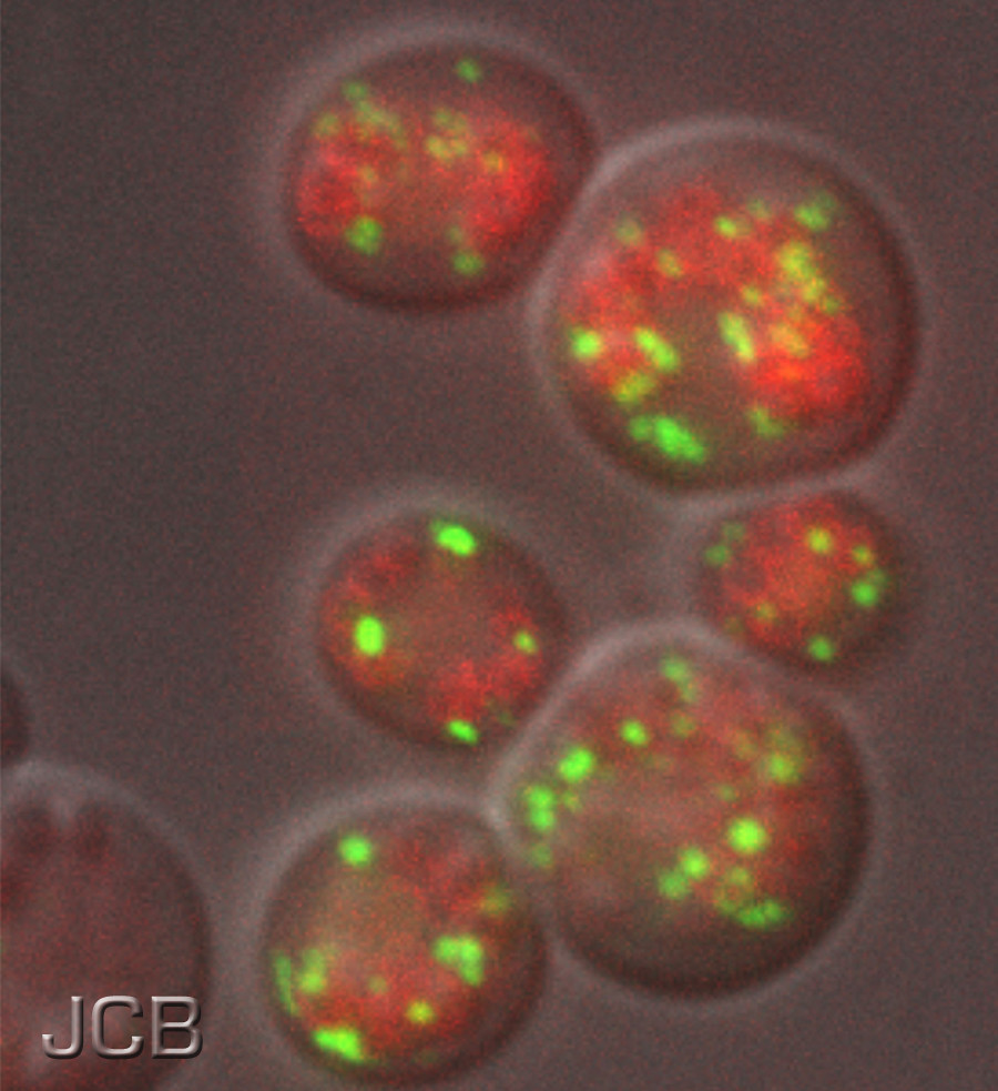
- Fluorescent stains like DAPI bind to DNA, making cell nuclei visible under UV light.
Using a Light Microscope
With your slide prepared and stained, you’re ready to use a light microscope. Start with a low magnification to locate your specimen, then gradually increase magnification for greater detail.
- Adjust the focus using the coarse and fine knobs.
- The objective lenses can be changed to increase magnification up to 1000x.
Electron Microscopy Insights
For even higher resolution, electron microscopy allows you to view structures at the nanometer scale. This technique uses beams of electrons instead of light to create an image.
- Transmission Electron Microscopy (TEM) offers insights into the internal structure of cells.
- Scanning Electron Microscopy (SEM) provides a detailed 3D view of cell surfaces.
Cellular Processes and Energy
Understanding how cells harness and utilize energy is fundamental to any animal project. The conversion and usage of energy within the cell allow for its growth, reproduction, and maintenance.
Photosynthesis vs. Cellular Respiration
Photosynthesis is a process unique to plant cells and some bacteria, where sunlight, carbon dioxide, and water are converted to glucose and oxygen. It’s an energy-storing process.
Cellular respiration, on the other hand, occurs in your animal cells. It’s the process by which cells take in oxygen and glucose to produce carbon dioxide, water, and most importantly, ATP, the energy that fuels cellular activities.
- Photosynthesis Equation: 6CO2 + 6H2O + light energy → C6H12O6 + 6O2
- Cellular Respiration Equation: C6H12O6 + 6O2 → 6CO2 + 6H2O + energy (ATP)
ATP: The Energy Currency of the Cell
ATP (Adenosine Triphosphate) is the energy currency of the cell. Think of ATP as a charged battery, ready to power cellular processes. When a phosphate group is removed, ATP becomes ADP (Adenosine Diphosphate), releasing energy for metabolic activities.
- Charged battery: ATP → Energy release → ADP + P + energy
Enzymes and Metabolic Pathways
Enzymes are proteins that accelerate, or catalyze, chemical reactions in your cells. Each step in a metabolic pathway typically requires a specific enzyme. These pathways are crucial as they guide the transformation of substances into energy or cellular building blocks. For instance, enzymes facilitate the breakdown of glucose during cellular respiration, resulting in the production of ATP.
- Enzyme action: Substrate → Enzyme catalysis → Products
- Example: Glucose → Glycolysis enzymes → Pyruvate (which enters other pathways to produce ATP)
Cell Cycle and Division
Understanding the cell cycle and division is fundamental in studying animal cells. You’ll explore how they replicate through mitosis, the role of meiosis in diversity, and the mechanisms controlling cell division.
Mitosis in Animal Cells
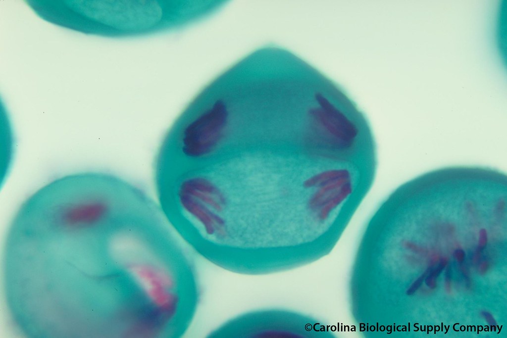
Mitosis is the process by which your animal cells divide, resulting in two genetically identical daughter cells.
It encompasses several phases: Prophase, where chromosomes condense and the nuclear envelope breaks down; Metaphase, where chromosomes line up at the cell’s equator; Anaphase, in which chromosomes separate to opposite ends of the cell; and Telophase, followed by Cytokinesis, where the cell splits into two. Teaching mitosis can involve hands-on activities to reinforce these concepts.
Meiosis and Genetic Diversity
Meiosis, unlike mitosis, results in four non-identical daughter cells, each with half the number of chromosomes of the original cell. This reduction is crucial for sexual reproduction, ensuring genetic diversity. During meiosis, crossover events increase diversity further by shuffling alleles between homologous chromosomes.
Regulation of Cell Cycle
The cell cycle is regulated by a series of checkpoints that ensure it is ready to proceed to the next phase. These checkpoints prevent mistakes, such as DNA replication errors, from being passed on.
Understanding these checkpoints further emphasizes the importance of cell cycle regulation in tissue health and the implications of their failure, which can lead to uncontrollable cell division, also known as cancer.
Cell Communication and Signaling
Cell communication and signaling are integral to understanding how cells in multicellular organisms behave and interact. These mechanisms are crucial for the processes of growth, development, and maintenance of homeostasis within an organism.
Hormones and Receptors
Hormones serve as messaging molecules that coordinate activities across different parts of your body. These chemical messengers bind to receptors, which are proteins located on cell surfaces. Upon binding, this interaction induces a specific response within the cell.
For example, the hormone insulin binds to insulin receptors, signaling cells to absorb glucose from the bloodstream.
Signal Transduction Pathways
Signal transduction pathways are complex series of events that translate extracellular signals into a functional response within the cell.
These pathways often begin with a signal molecule (such as a hormone) binding to a receptor, leading to a cascade of molecular interactions, and ultimately resulting in changes in gene expression or cellular activity.
An example of signal transduction is the pathway initiated by epidermal growth factor (EGF), which promotes cell division.
Neurotransmitters and Synaptic Transmission
Neurotransmitters are chemicals used by neurons to communicate with each other or with other types of cells. They are released from neurons in response to electrical signals, cross the synaptic gap, and bind to receptors on the target cell.
This binding triggers a change in the target cell, for instance, propagating an electrical signal or causing muscle contraction. A well-known neurotransmitter is acetylcholine, which plays a role in muscle movement and memory.
Animal Cell Culturing Techniques
Mastering the fundamentals of animal cell culturing is vital for any laboratory work related to cell science. Accurate techniques ensure cell viability and the success of downstream applications.
Creating a Sterile Environment
To establish a sterile environment, you must work within a laminar flow hood which provides a contamination-free workspace. Surfaces should be regularly disinfected with ethanol or other suitable germicides, and all media, reagents, and equipment must be sterilized before use, often through autoclaving or filtration to eliminate the risk of contamination.
Cell Line Selection and Maintenance
Selecting the right cell line for your project is pivotal. Each line has specific growth requirements and characteristics, such as growth rates and morphology.
Once selected, maintaining your line requires precise culture conditions including optimal temperature, usually at 37°C, and a CO₂ atmosphere to maintain the correct pH.
Regular subculturing is necessary to keep the cells healthy and prevent over-confluence which can negatively affect cell behavior and viability.
Assaying Cell Vitality and Proliferation
Quantifying cell health and proliferation involves several methods. MTT assays, for example, allow you to assess cell metabolic activity whereas Trypan Blue exclusion tests provide a direct count of viable cells. It’s crucial to perform these assays regularly to monitor cell health, especially before conducting any experiments that depend on cell viability and growth characteristics.
Application of Cell Science in Medicine
In medicine, cell science plays a pivotal role, particularly in advancing drug development, fostering regenerative therapies, and enhancing cancer treatments. Your understanding of these applications is crucial in grasping the potential and progress in modern medicine.
Drug Development and Testing
You will find that cell-based assays are integral to pharmaceutical innovation. Using animal cells, researchers can test the efficacy and safety of new compounds. This process significantly reduces the time and resources required to identify potential drugs before they proceed to clinical trials.
For instance, monoclonal antibodies are produced using hybridoma technology, leading to groundbreaking treatments for diseases like cancer and autoimmune disorders.
Stem Cells and Regenerative Medicine
Stem cell technology holds a transformative promise for regenerative medicine, where your body’s own cells can heal and replace damaged tissues. Mesenchymal stem cells (MSCs), with their regenerative and immunomodulatory properties, are a cornerstone of current therapy advances in both human and veterinary medicine.
Cancer Cells and Oncology
Your awareness of cancer treatment progresses hand-in-hand with innovations in cell science. Cultured cancer cells allow for in-depth study of tumor biology and the development of targeted therapies.
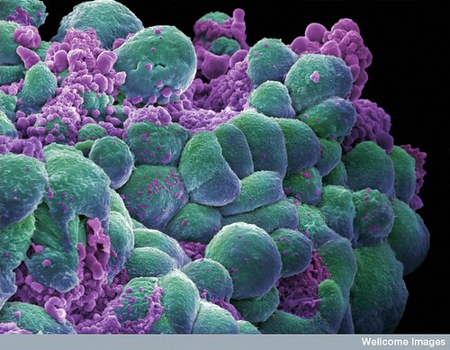
These studies facilitate personalized medicine approaches, making treatments more effective and less invasive. Furthermore, understanding how cancer cells evade the immune system paves the way for novel immunotherapies.
Biotechnology in Animal Cell Science
Biotechnology harnesses cellular and biomolecular processes to develop technologies that enhance our understanding and manipulation of animal cells. The advent of sophisticated tools and methodologies has revolutionized this field, offering profound applications in medicine, research, and industry.
Genetic Engineering
Your exploration of biotechnology in animal cell science begins with genetic engineering, a cornerstone of modern biology. Through techniques like recombinant DNA technology, you can introduce new genetic material into animal cells, leading to the expression of desirable traits or production of biologics.
For instance, the production of high-value bioproducts such as antibodies, which rely on animal cells due to their glycosylation capabilities, exemplifies the advancements in this domain.
Protein Synthesis and Purification
By leveraging animal cell systems, you have the capacity to synthesize complex proteins that cannot be adequately produced in simple microbial systems. Protein purification is the subsequent, critical step that isolates these proteins in their pure form for various applications, ranging from therapeutic use to research tools.
The field has evolved to develop more refined purification techniques, ensuring the proteins fulfill their potential in applications where structural integrity and functionality are paramount.
CRISPR and Genome Editing
At the forefront of genome editing is CRISPR-Cas9, a revolutionary technology that allows for precise, direct modification of the genome within living organisms. It has been instrumental in editing genes in animal cells, with applications in creating disease models for research and potential therapies for genetic disorders.
Advancements made by researchers, such as developing methods to genetically modify individual cells in animals, have opened new avenues for in-depth biological studies and therapeutic interventions.
Science Project Design and Execution
In executing your animal cell science project, it’s vital to systematically approach each stage from hypothesis formulation to presenting your findings. Detailed planning and adherence to scientific methods are the cornerstones of a successful project.
Formulating a Research Question
Your science project begins with a research question. Determine what you aim to discover about animal cells, such as the effect of various substances on cell health. This question should be specific, measurable, attainable, relevant, and time-bound (SMART).
Experiment Design and Control Groups
Design your experiment with clear independent and dependent variables. If investigating a cell component’s function, establish control groups to provide reliable data for comparison. Use materials like clay or a gelatin model to represent the parts, ensuring each variable is controlled to isolate your experiment’s focus.
Data Collection and Analysis
Gather data methodically during your experiment to ensure accuracy. Record measurements and observations in an organized manner, possibly using a table format for clarity. Afterward, employ appropriate statistical analysis tools to discern patterns or anomalies in your results.
Presenting Scientific Findings
Finally, present your findings with certainty. Use visual aids like charts or a 3D cell model to illustrate your data, and be prepared to explain the significance and the scientific concepts that your project addresses. Your presentation should be clear, concise, and substantiated by your gathered data.
Frequently Asked Questions about Animal Cell Science Projects
In this section, you’ll find clear, concise answers to common queries on creating animal cell science projects. This practical guide aims to help you successfully construct and understand your project by addressing material needs, labeling techniques, edible project options, cell types, creative project ideas, and the distinguishing features between animal and plant cells.
What materials are needed to construct a 3D animal cell model?
To create a 3D animal cell model, you will need modeling materials such as clay, playdough, or gelatin. Objects like beads, buttons, and yarn can serve as organelles. For a more detailed overview, explore DIY Science Project Ideas – Osmo.
How can I label the organelles in an animal cell project?
Labels can be made using small pieces of paper and toothpicks or by directly writing on the model if the material allows. For a non-permanent option, use sticky notes or flags positioned beside each organelle.
What edible items can represent organelles in an animal cell cake project?
Candies and frosting are excellent for representing organelles on an animal cell cake. Fruit slices, sprinkles, and various colored icing can illustrate parts like the nucleus, mitochondria, and ribosomes. For inspiration, check out How to Make an Animal Cell for a Science Project | Sciencing.
Can you list five types of animal cells commonly studied in biology?
Commonly studied animal cells in biology include muscle cells, nerve cells, blood cells, epithelial cells, and fat cells. Each type plays specific roles in the body’s structure and function.
What are some creative ideas for a 6th-grade animal cell project?
For a 6th-grade animal cell project, consider making a cell model with household items, constructing a cell from cake or gelatin, or creating a digital 3D representation.
Encourage creativity by using various colors and textures to depict different organelles. Find creative project ideas on Easy & Fun Animal Cell Science Projects for Kids – Homeschool Giveaways.
How does an animal cell differ from a plant cell in structure and function?
Animal cells differ from plant cells by lacking a cell wall and chloroplasts, and typically having smaller vacuoles. Functionally, animal cells do not perform photosynthesis and have different methods for storing energy and maintaining structure.

Leave a Reply